


« “El PET permite encontrar mucho antes la lesión tumoral”
Hodgkin’s lymphoma, PET scan
El tamaño completo es de 530 × 506 pixels
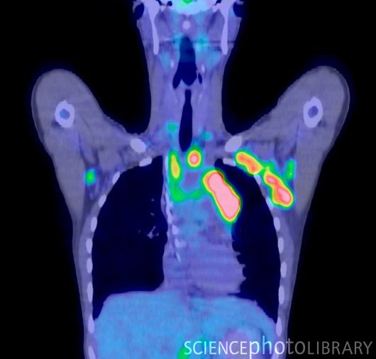
Hodgkin’s lymphoma. Image 1 of 2. Combined computed tomography (CT) and positron emission tomography (PET) scan of cancerous sites (coloured areas) in a patient with Hodgkin’s lymphoma (cancer of the lymph nodes). This is a coronal scan, passing vertically through the body seen from the front. The cancer is present in the bilateral and mediastinal axillar lymph nodes. This is 2-18F-fluoro-2-deoxy-D-glucose (FDG) positron emission tomography. FDG is a radioactive tracer that can be used to identify cancer sites. For the same patient scanned after treatment by chemotherapy, see image C001/7582.